Our human temporal bone collections has the largest number of otitis media cases worldwide. Our collection of otitis media cases have been used by hundreds of fellows and researchers in the past. These temporal bones were used in groundbreaking studies that have changed paradigms regarding otitis media. For example, Dr. Paparella published, in the 1970s, the first study corroborating that otitis media can result in sensorineural hearing loss. Our laboratory also published a number of studies proving that the round window membrane is the portal through where toxins and bacterial products can penetrate to the inner ear, causing loss of hair cells. Additionally, Dr. Paparella also described the "continuum of otitis media", demonstrating the series of events leading to progression of otitis media to a chronic disease. Representative slides from our collection are below:
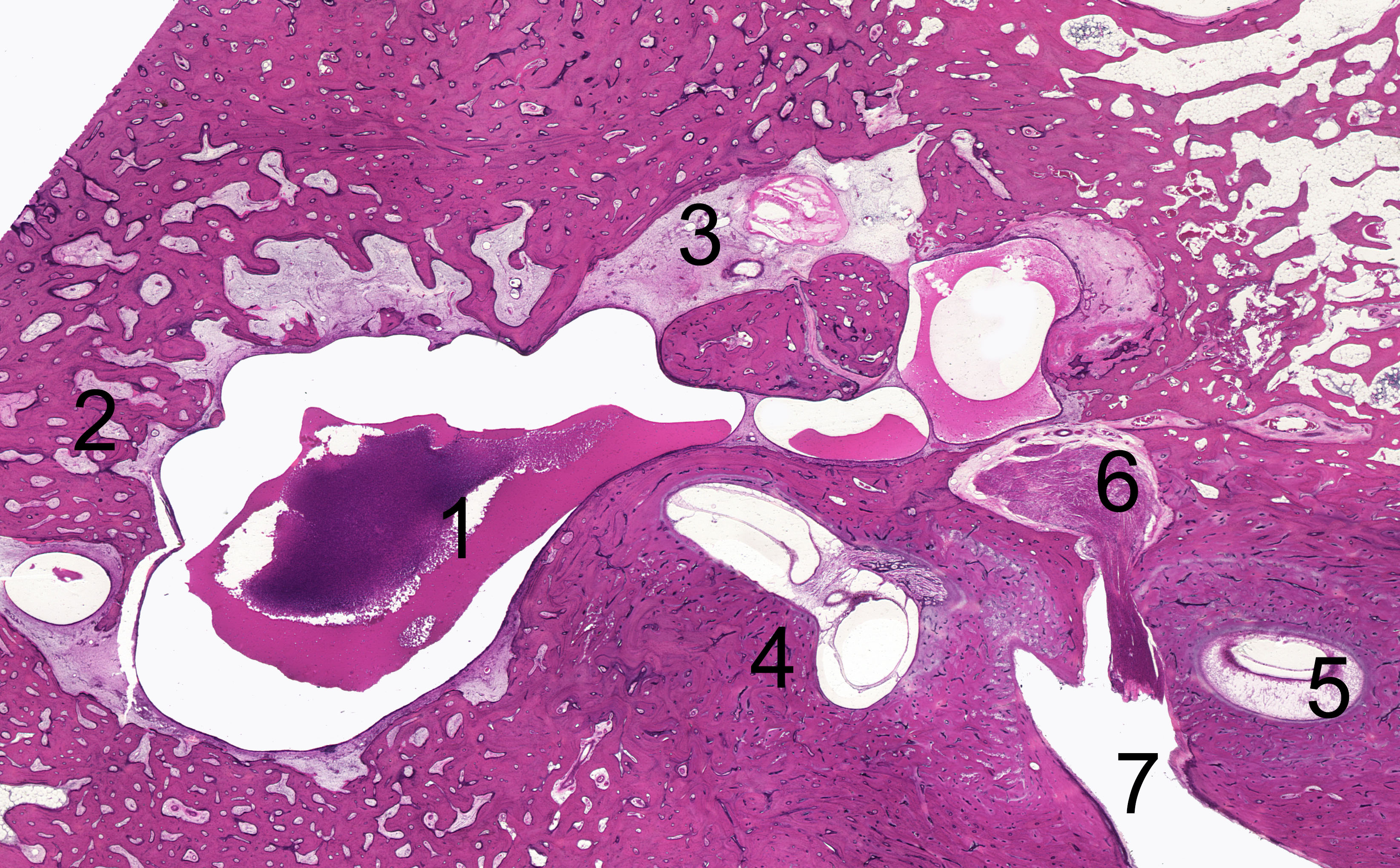
Figure 1. A representative horizontal human temporal bone section from a patient with chronic otitis media (COM). The mastoid antrum and middle ear are filled with mucoid-purulent effusion (1). The mastoid is sclerotic (2), and fibrous tissue can be seen in the anterior aspect of the malleus and incus (3). (4) = Lateral semicircular canal; (5) = cochlea; (6) First genu of the facial nerve; (7) Internal auditory canal
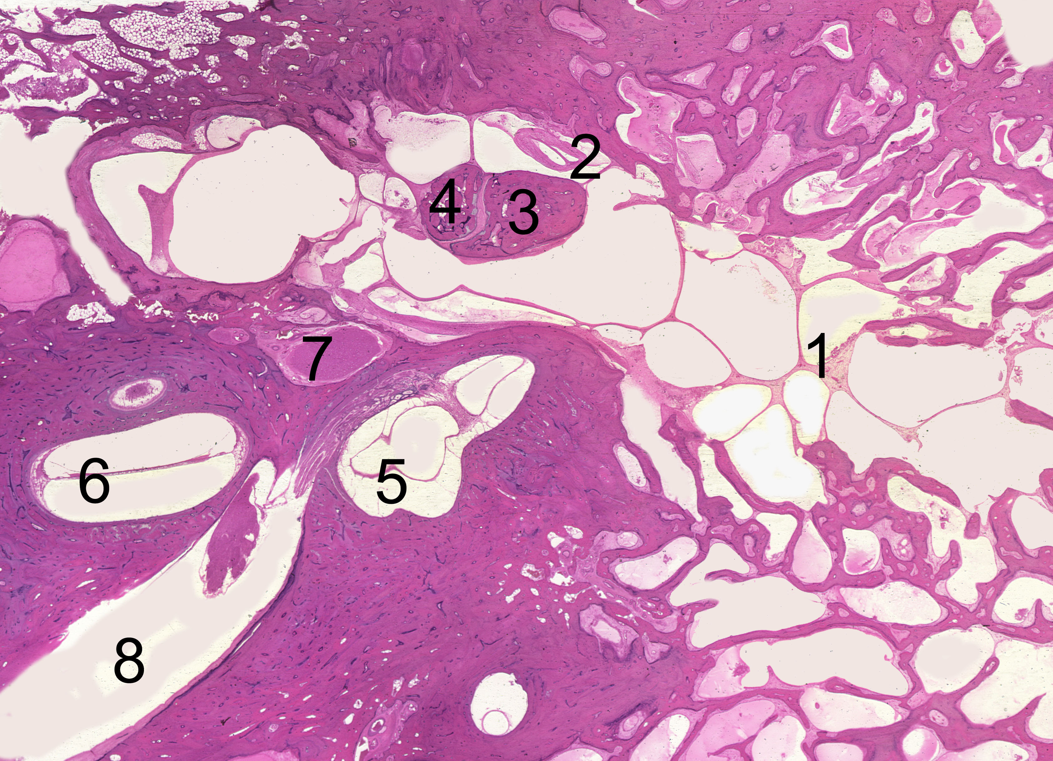
Figure 2. A representative human temporal bone horizontal section from a patient with chronic otitis media stained in H&E. The picture shows the presence of serous effusion, fibrous tags, and granulation tissue in a well-pneumatized mastoid (1). (2) = cholesterol granuloma in the anterior epitympanic compartment of the middle ear. (3) = Short process of the incus; (4) = Malleus head; (5) Lateral semicircular canal; (6) Cochlea; (7) Tympanic portion of the facial nerve; (8) Internal auditory canal.