Processing services and temporal bone tissue we provide
The otopathology laboratory at the University of Minnesota has multiple resources to facilitate collaborative research efforts with nationwide (and international) researchers. Our laboratory houses over 2,200 human temporal bone specimens that have been amassed through decades. The collection includes specimens with virtually every disease and disorder that affect the human hearing and balance, including 16 genetic diseases and numerous fetal and pediatric bones. We also have ~800,000 unstained sections that can be reprocessed and used for a variety of purposes (e.g., use of different stains, immunohistochemistry protocols, and tests for retrieving proteins and genetic material). We have a large procurement capacity of temporal bones (up to 400/year) that affords our capacity to use specimens for experimental approaches, different types of processing and sectioning, as well as proposing and implementing innovative methods for temporal bone studies.
How to request processing services or tissue
To request processing services and tissue, please use our tissue request form. Alternatively, please submit the following information to the email [email protected]:
1) A description of the project’s purpose, how tissues will be utilized, required tissue & preservation criteria, if known; and
2) PI’s CV/biosketch
Our team will work with requestors and the PI/staff to approve and refine requests (all responses within 2 business days) to best meet study needs while complying with best practices. The requests will be analyzed by a panel comprised by the PIs (Drs. Cureoglu and Adams) and the research staff (Dr. Monsanto and Nevra Keskin). Priority will be given to feasible studies aiming to answer well-defined research questions foundational to otologic- or neuroscience, translation of animal models to humans, and assessment or refinement of clinical therapies and procedures. If feasibility of the proposed investigation is assessed, a meeting will be coordinated between the requestors and our laboratory staff members to further discuss specific timeline and logistics of the experiments.
Fresh tissue can be acquired with a short turnaround time and processed according to the researchers needs. For these requests, we will work with ABP to prioritize procurement of specimens of special scientific value to the research (e.g., diverse, short postmortem time, functional auditory/vestibular testing, specific health conditions/surgeries). Standard processing of freshly acquired temporal bones can take up to 2 years, but specific temporal bones that are currently being processed can be prioritized, whether from donors with no history of ear diseases (“controls”) or those with identified conditions. Whole mount sectioning can be done after the temporal bone is decalcified. Stained archival tissue can be made immediately available through slide scanning. Our archival collection also contains ~800,000 unstained sections that can be de-celloidinized and used for staining, application of histochemistry, and immunohistochemistry.
We can distribute processed tissue nationwide (if applicable) through the mailing services provided by UMN. We can ship biological material (non-infected and infected) that is immersed or embedded in chemicals. The shipping costs will be charged to researchers requesting the tissues.
A list of the processing services our laboratory provides include (but is not limited to):
| Service | Description/options |
| Full processing | We can fully process temporal bones (animals and humans) harvested by external scientists |
| Standard processing | Formalin fixation, decalcification, celloidin-embedding, staining with H&E |
| Decalcification | Ethylenediaminetetraacetic acid, microwave decalcification |
| Embedding | Celloidin, paraffin, polyester wax, frozen, epoxy |
| Sectioning | Horizontal, vertical, cryostat, ultratome |
| Microdissection | Whole-mount and surface preparation sections of endorgans |
| Preparation for microscopy | Light, differential interference contrast, fluorescence, confocal, electron |
| Staining | H&E, different stains, histochemical, immunohistochemistry, in-situ hybridization |
| Re-processing of unstained sections | Archival sections that can be re-processed, stained, and used for histochemistry and immunohistochemistry and immunohistochemistry (optimal results: specimens processed <10 years ago) |
| Non-destructive imaging techniques | Thin-sheet laser imaging microscopy; light-sheet |
| 3-D reconstructions | Using scanned, stained slides, through AMIRA software (available at the University of Minnesota Imaging Centers for an hourly fee) |
| Digitization of stained slides | Using a high-resolution, high-throughput scanner (5-40x of magnification) |
| Validation studies (animals-humans) | Through processing human specimens similarly to our animal collection |
Facilities
The Otopathology Laboratory at the University of Minnesota offers a variety of methods for procurement, processing, and analysis of human temporal bones. In our facility, we have 2000+ archival human temporal bone specimens that are ready for multiple analyzes, including light microscopy and three-dimensional reconstructions.
We have a wet laboratory that contains the necessary materials and equipment to process human and animal temporal bones. Our animal housing facilities has full-time personnel and veterinarians on call to allowing optimal care of the animals used in our studies. We have access to numerous microscopes and imaging devices through our own laboratory and research partners, including light and electron microscopes, thin light-sheet microscopes, fluorescence and confocal microscopes, high-resolution scanners and 3-dimensional reconstruction softwares, high-resolution CT scans, and the world's highest resolution MRI equipment (16.5T).
University Imaging Centers
The University Imaging Centers (UIC) is a network of shared research facilities located on the University of Minnesota-Twin Cities campuses: Snyder Hall on the St. Paul campus, Jackson Hall on the Minneapolis East Bank campus, and the Cancer and Cardiovascular Research Building located in the Biomedical Discovery District on the East Bank campus.
The UIC constitutes a national center of excellence of tissue processing and cutting-edge imaging devices. It offers equipment and support for light and electron microscopy, multiplex ion beam imaging, tissue clearing, in-vivo imaging from subcellular to whole animals, image analysis & visualization, slide scanning, large format poster printing, and 3D printing. Services range from training users to independently operate equipment to full-service imaging, and sample processing. Operating under the direction of Mark Sanders, the UIC has seven full-time staff members, three part-time, as well as multiple undergraduate employees that can assist in experimental design, probe and labeling selection, sample handling, tissue clearing, as well as data analysis and interpretation.
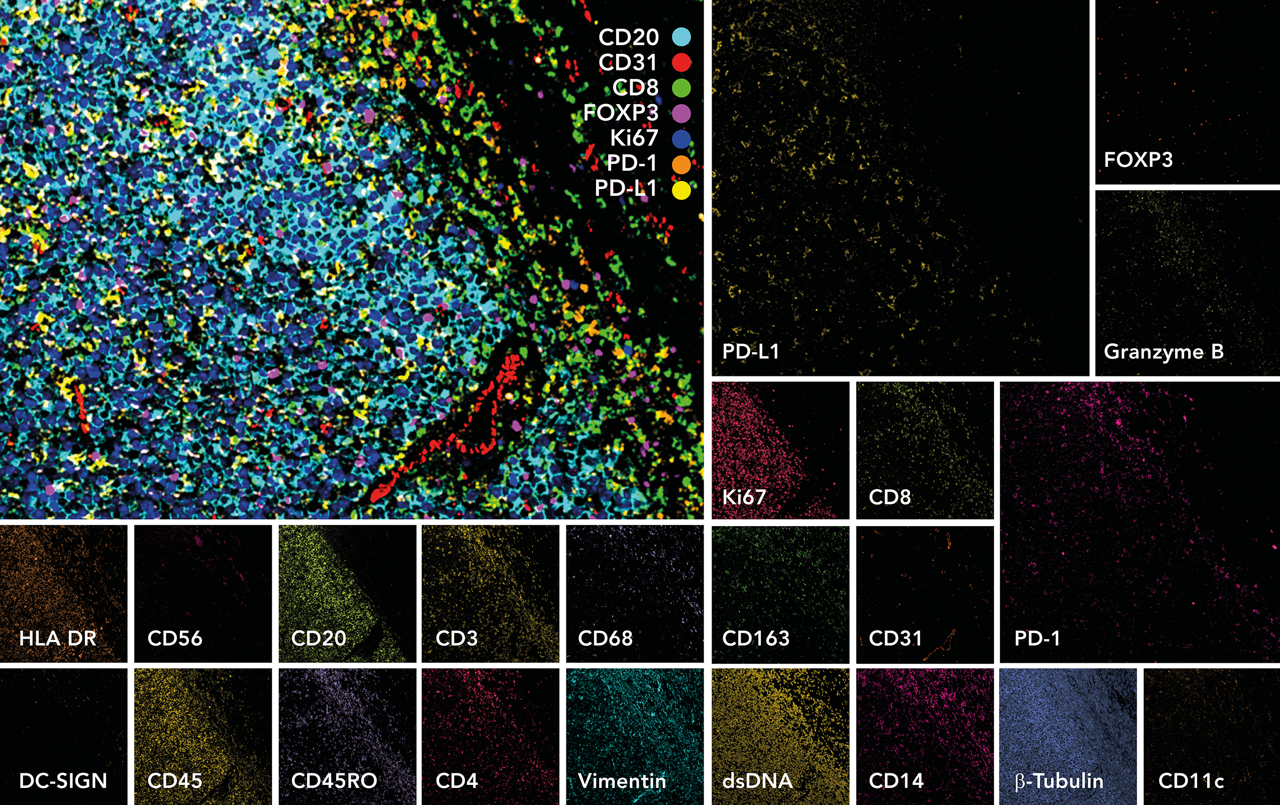
The UIC currently serves 53 different departments within the University of Minnesota (241 investigators), having 522 active equipment users. Their resources include (but are not limited to) two laser scanning confocal microscopes with multiphoton capabilities, six single photon confocal microscopes (three with spectral unmixing), super resolution structured illumination microscope (SIM), spinning disk confocal microscope, total internal reflection fluorescence (TIRF) microscope, fluorescent spectral meso-confocal microscope, automated wide-field microscopes, high-speed ribbon confocal microscope, and light-sheet microscope.
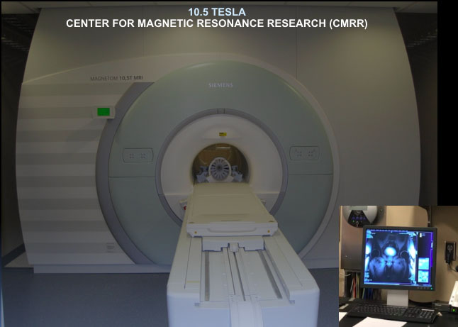
Center for Magnetic Resonance Research
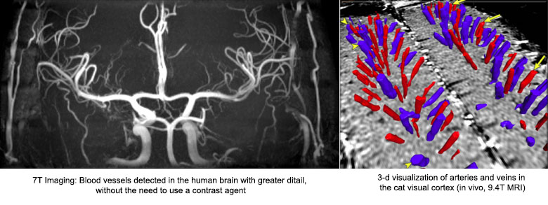
Center for Magnetic Resonance Research (CMRR), funded as a Biotechnology Research Center (BTRC) by the National Center for Research Resources (NCRR) until 2012 and by the National Institute of Biomedical Imaging and Bioengineering (NIBIB) since then, focuses on development of unique magnetic resonance (MR) imaging and spectroscopy methodologies and instrumentation for the acquisition of structural, functional, and biochemical information non-invasively in humans, and utilizing this capability to investigate organ function in health and disease. The distinctive feature of this center is the emphasis on ultrahigh magnetic fields (7 Tesla and above), which was pioneered by this BTRC. The CMRR houses the world's highest resolution whole-body MRI (10.5T) available for use in humans, and also the world's highest resolution research MRI (16.5T).
This emphasis is based on the premise that there exists significant advantages to extracting biomedical information using ultrahigh magnetic fields, provided difficulties encountered by working at high frequencies corresponding to such high field strengths can be overcome by methodological and engineering solutions. CMRR is home to some of the most advanced MR instrumentation in the world, complemented by human resources that provide unique expertise in imaging physics, engineering, and signal processing.
No single group of scientists can successfully carry out all aspects of this type of interdisciplinary biomedical research; by bringing together these multi-disciplinary capabilities in a synergistic fashion, facilitating these interdisciplinary interactions, and providing adequate and centralized support for them under a central umbrella, CMRR amplifies the contributions of each of these groups of scientists to basic and clinical biomedical research. Collectively, the approaches and instrumentation developed in CMRR constitute some of the most important tools used today to study system level organ function and physiology in humans for basic and translational research and are increasingly applied world-wide.